PIE-RICS and RLICS
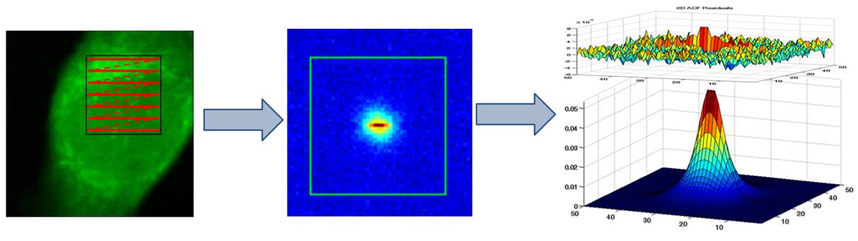
For raster image correlation spectroscopy (RICS) images are recorded by rapidly scanning a small area of the sample with a laser scanning confocal microscope. Due to the acquisition via raster scanning, there is not only a spatial but also a temporal dependence between the pixels. Thus diffusion coefficients can be extracted from the spatial correlation of the images by using a special fit. In contrast to FCS, RICS allows one to fit both focus size and diffusion coefficient simultaneously, making it a calibration free method. Furthermore, immobile or slow moving contributions can be filtered out quite easily and the rapid scanning reduces bleaching. This makes RICS ideal for measurements in vivo and for fluorescent proteins. As for point FCS, cross-talk free cross-correlations are achieved by combining RICS with PIE. Additionally, different species can be distinguished by their fluorescence lifetime using raster lifetime image correlation (RLICS).
- Brown, C. M., et al. "Raster image correlation spectroscopy (RICS) for measuring fast protein dynamics and concentrations with a commercial laser scanning confocal microscope." Journal of microscopy 229.1 (2008): 78-91.
- Hendrix, Jelle, et al. "Pulsed interleaved excitation fluctuation imaging."Biophysical journal 105.4 (2013): 848-861.