Chemical Biology of Protein Post-Translational Modifications
Neurological disorders and in particular neurodegeneration is becoming ubiquitous in ageing populations. Regardless of many efforts, the fundamental principles underlying the mechanisms of failing neurodevelopment and onset of the neurodegeneration are still unknown. The protein post-translational modifications (PTMs) are closely connected to multiple metabolic and signalling pathways involved in neurodegeneration.
Our research focuses on development of chemical biology tools to analyse protein post-translational modifications. We combine induced pluripotent stem cells-based cellular models, quatitative mass spectrometry techniques and organic synthesis to track and visualize PTMs in living cells.
Protein AMPylation
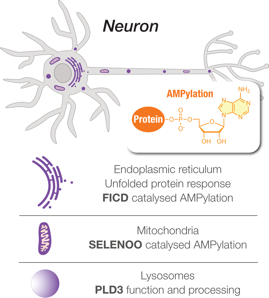
Protein AMPylation is a post-translational modification in which an adenosine monophosphate (AMP) moiety from ATP is covalently attached to the side chain of a serine, threonine or tyrosine residue. In humans, only two AMP-transferases are known so far: FICD, which is localised to the endoplasmic reticulum, and SELENOO, which is localised to mitochondria. Despite its emerging role in protein homeostasis and neuronal development, the full scope of AMPylated proteins and the functional consequences of this modification remain largely unexplored.
Recently, our group has identified the Alzheimer's disease-associated protein PLD3 as specifically AMPylated in the lysosomal lumen of young and mature neurons. We have established a novel gel-based methodology that separates the modified and unmodified forms of proteins, complementing our mass spectrometry data. Together, these approaches have revealed a fascinating processing pathway of PLD3 during neuronal differentiation and maturation. Moreover, using data-independent acquisition (DIA) mass spectrometry, we have identified the AMPylation sites of PLD3 and shown that without them, PLD3 is no longer proteolytically processed, which largely inhibits its exonuclease activity.
Selected publications:
Koler et al. bioRxiv 2026 DOI: 10.64898/2026.03.30.715195.
Hoffmann et al. Mol. Cell. Proteomics 2025 DOI: 10.1016/j.mcpro.2025.101051.
Becker et al. iScience 2021 DOI: 10.1016/j.isci.2021.103521.
Sieber et al. Cell Chem. Biol. 2020 DOI: 10.1016/j.chembiol.2020.05.009.
Kielkowski et al. Nat. Commun. 2020 DOI: 10.1038/s41467-019-14235-6.
Tubulin Code
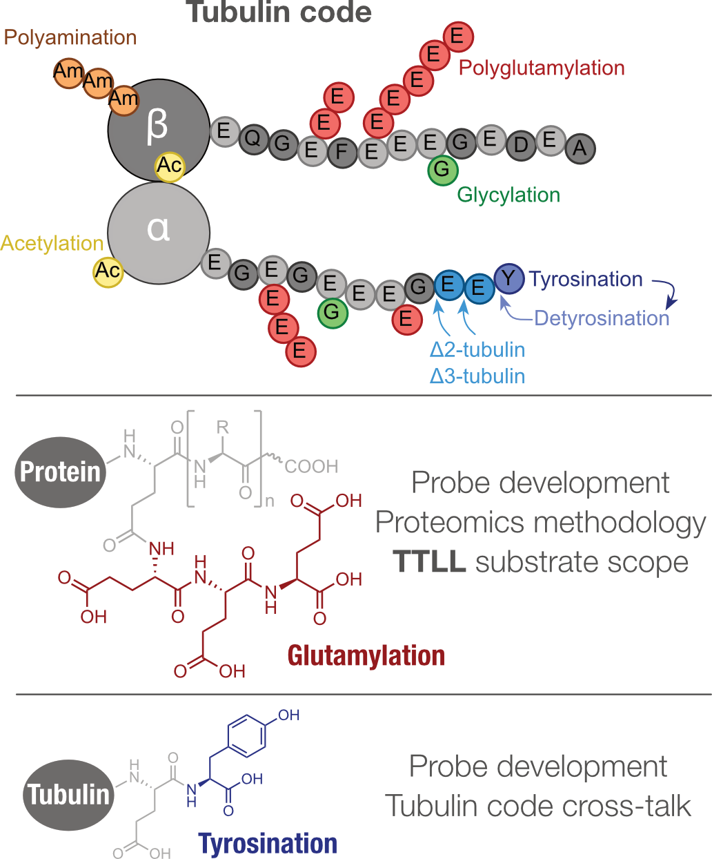
Microtubules are decorated with a variety of post-translational modifications, collectively referred to as the tubulin code, that fine-tune their stability and their interactions with motor and microtubule-associated proteins. Most of these modifications are catalysed by the tubulin tyrosine ligase-like (TTLL) family, which comprises 13 members in mammals. Nine of them are glutamylases, divided into initiating mono-glutamylases and elongating poly-glutamylases, while others catalyse glycylation. In contrast, tyrosination, the re-addition of a C-terminal tyrosine to detyrosinated tubulin, is catalysed by a single enzyme, tubulin tyrosine ligase (TTL), and has so far been described exclusively on alpha-tubulin. Despite their importance, these modifications remain understudied, even though dysregulation of several tubulin code enzymes has been linked to neurodegeneration, cancer and retinitis pigmentosa.
To study protein tyrosination directly in living cells, we have established an O-propargyl-L-tyrosine probe that exploits the substrate promiscuity of TTL and enables mass spectrometry-based chemical proteomic profiling of tyrosinated proteins. Using this approach, we found that the alpha-tubulin isotype TUBA4A also undergoes tyrosination. This was unexpected, because the TUBA4A gene does not encode a C-terminal tyrosine, so it was previously not thought to be tyrosinated. The finding is particularly interesting because TUBA4A is a neuron-enriched tubulin isotype, which suggests a specific role of tyrosination in fine-tuning the neuronal microtubule network.
Selected publications:
Makarov et al. ChemBioChem 2022 DOI: 10.1002/cbic.202200414.
Chemical Proteomics Methodology
To analyse protein PTMs, we use mass spectrometry-based chemical proteomics. Given the growing evidence that PTMs play a crucial role in neurological disorders, we develop chemical proteomic strategies for the unambiguous analysis of PTMs in the different cell types of the central nervous system, and we continuously improve every step of the workflow.
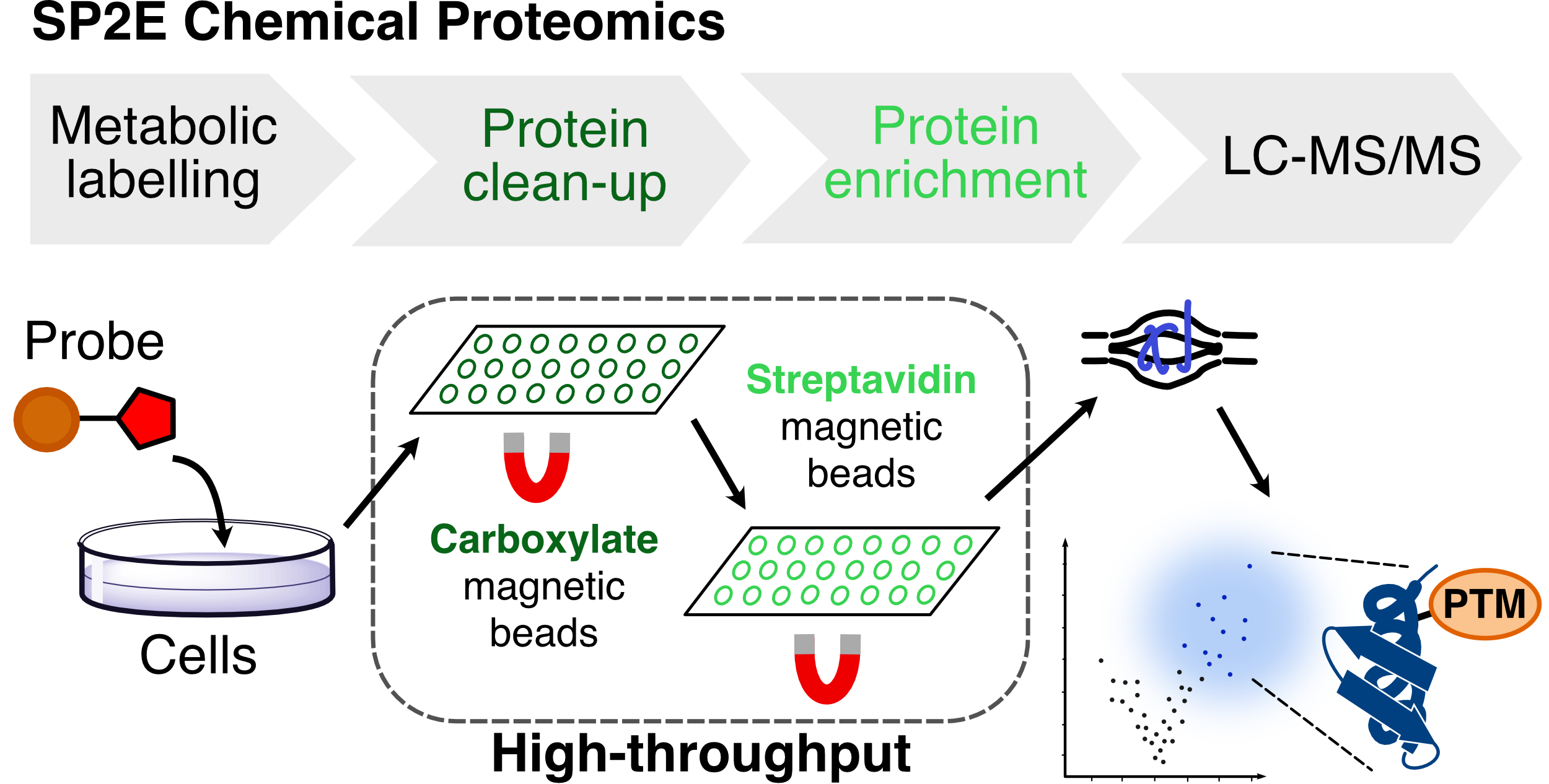
To streamline the enrichment of probe-labelled proteins, we developed the SP2E workflow, which combines protein clean-up on carboxylate-coated magnetic beads with enrichment on streptavidin-coated magnetic beads. The workflow is robust and reproducible and runs in a 96-well plate format suitable for automation. Because it requires only small amounts of protein input, it is particularly well suited to precious samples such as human iPSC-derived neurons, which are demanding and costly to culture.
We also systematically tested the copper-catalysed azide-alkyne cycloaddition (CuAAC), the 'click reaction' at the heart of most chemical proteomic experiments. We discovered that, under standard conditions, free cysteine thiols react with the alkyne probe and the azide tag to form thiotriazole adducts. These adducts create a ubiquitous background and false-positive hits. We showed that this side reaction can be suppressed by a simple adjustment of the protocol, increasing the concentration of the reducing agent TCEP, which markedly improves the signal-to-noise ratio. Both SP2E and the improved click conditions have been widely adopted by the chemical proteomics community.
Selected publications:
Abanti et al. RSC Chem. Biol. 2026 DOI: 10.1039/d6cb00004e.
Chen et al. Chem. Sci. 2025 DOI: 10.1039/d5sc00400d.
Wiest et al. JACS 2024 DOI: 10.1021/jacs.3c11780.
Becker et al. JACS Au 2022 DOI: 10.1021/jacsau.2c00284.
Makarov et al. J. Mass Spectrom. 2022 DOI: 10.1002/jms.4812.